Evaluating the Elbow
|
|
|
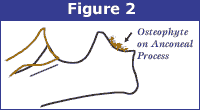
Schematic representation of the radius and ulna of a normal elbow joint. Note the smooth margin of the anconeal process of the ulna (angled line) |
Elbow dysplasia has multiple inherited etiologies which may occur singularly or in combination. These etiologies include fragmented medial coronoid (FCP) of the ulna, osteochondritis of the medial humeral condyle and ununited anconeal process (UAP). The most sensitive view used to diagnose secondary degenerative changes in the elbow joint is an extreme flexed medio-lateral view of the elbow (Figure 1) which is required by the OFA and recommended by the International Elbow Working Group. The veterinary radiologists are most interested in the appearance of the anconeal process of the ulna.
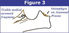
When there is instability of the elbow joint due to elbow dysplasia, one of the most sensitive radiographic findings is new bone proliferation (osteophytes) on the anconeal process of the ulna (Figure 2) associated with secondary developmental degenerative joint disease.

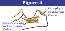
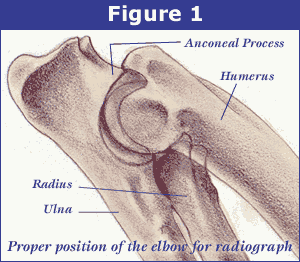
Bone proliferation can be very subtle to visualize in some dogs and may require the use of a special light source (hot light) rather than a traditional view box to diagnose it. Other arthritic findings such as sclerosis in the area of the trochlear notch of the ulna and bone spurs at joint edges are also reported. If fragmentation of the medial coronoid only involves the cartilage, it may not be seen radiographically but occasionally if the bone is also fragmented, it can be visualized as a separate calcific opacity superimposed over the radius (Figures 3 and 4).


Grading Elbows
For elbow evaluations, there are no grades for a radiographically normal elbow. The only grades involved are for abnormal elbows with radiographic changes associated with secondary degenerative joint disease. Like the hip certification, the OFA will not certify a normal elbow until the dog is 2 years of age. The OFA also accepts preliminary elbow radiographs. To date, there are no long term studies for preliminary elbow examinations like there are for hips, however, preliminary screening for elbows along with hips can also provide valuable information to the breeder.
Grade I Elbow Dysplasia
Minimal bone change along anconeal process of ulna (less than 3mm).
Grade II Elbow Dysplasia
Additional bone proliferation along anconeal process (3-5 mm) and subchondral bone changes (trochlear notch sclerosis).
Grade III Elbow Dysplasia
Well developed degenerative joint disease with bone proliferation along anconeal process being greater than than 5 mm.
